
Introduction
Platelets are the second most abundant cellular component of blood, after erythrocytes, with a circulating half-life of 7 to 10 days. They are anucleated cellular fragments derived from bone marrow megakaryocytes, and in healthy humans their size ranges between 2 and 5mm. Platelets are prepackaged with proteins and bioactive molecules that allow them to respond rapidly to stimuli without the need for activation of transcription or translation processes. These molecules are stored in three main types of granules: a granules (containing proteins such as growth factors, chemokines, adhesion molecules), dense or d granules (with serotonin, ATP, ADP and Ca²+) and lysosomes (with specific hydrolytic enzymes)1 (Tables 1 and 2).
Table 1. Characteristics of Alfa granule
| Categories | Alpha granule content |
|---|---|
| Adhesive proteins | TSP-1, TSP-2, laminin-8 |
| Coagulation factors | Factor V/Va, Factor XI, multimerin, protein S, HMWK, protease, nexin-1 and 2, TFPI, protein C inhibitor |
| Fibrinolytic factors | Plasminogen, PAI-1, α2-antiplasmin, TAFI, α2 macroglobulin |
| Proteases and antiproteases | MMP-1,23,4,9, ADAMTS13, ADAMS10, ADAMS17, TIMPs 1-4, platelet inhibitor of FIX, C1 inhibitor, α1 antitrypsin |
| Growth factors | PDGF, IGF-1, VEGF (A and C), CTGF, IGFBP3 |
| Chemokines, cytokines and others | TGF B1 and B2, IL-1, IL-8, PF-4, NAP-2, angiopoietin-1, endostatin, osteoprotegerin |
| Antimicrobial proteins | Thrombocidins, quinocidins |
| Membrane glycoproteins | αIIbβ3, αvβ3, PECAM-1, receptors for primary agonists, P-selectin, TLT-1, TF, furin, cellubrevina, syntaxin-2, clathrin |
|
TSP-1 and 2: thrombospondin; Factor V: proaccelerin or labile factor; Factor XI: plasma thromboplastin antecedent; TFPI: tissue factor pathway inhibitor; PAI-1: tissue-type plasminogen activator inhibitor type 1; TAFI: thrombin-activatable fibrinolysis inhibitor; MMP: metalloproteinases; ADAMTS10-13: a disintegrin and metalloprotease with thrombospondin type 1 motifs num. 10, 13 and 17; TIMP: tissue inhibitors of metalloproteinases; FIX: antihemophilic factor B; PDGF: platelet-derived growth factor; IGF: platelet-derived growth factor; VEGF: vascular growth factor; CTGF: connective tissue growth factor; IGFBP3: insulin-like growth factor-binding protein 3; TGF B1 and B2: transforming growth factor; IL: interleukin; PF: platelet factor; NAP-2: 2-thromboglobulin-F; αIIbβ3: GP IIb-IIIa complex; αvβ3: vitronectin receptor; PECAM-1: platelet endothelial cell adhesion molecule 1; TLT-1: TREM-like transcript 1. |
|
Table 2. Characteristics of dense granules
| Categories | Dense granule content |
|---|---|
| Cell signaling | N-terminal domain inhibitor of calmodulin, 14-3-3 protein |
| Chaperone molecules | Cyclophilin A, heat shock protein 70 kDa, disulfide-isomerase |
| Cytoskeleton | B actin, α1 actin, adenylate cyclase-associated protein, cofilin 1, filamin A, myosin light chain-2, pleckstrin, talin-1, thrombopoietin chain b, tropomyosin, vinculin |
| Glycolysis | Aldolase, α-enolase 1, GAPDH, lactate dehydrogenase B, pyruvate kinase |
| Related to platelet function | Beta-thromboglobulin precursor, FXII, fibrinogen alpha chain precursor, Ig gamma chain C region, platelet factor 4, glycoprotein 11, pro-platelet precursors, serum albumin, thromboplastin-1 precursor |
|
Heat shock protein 70 kDa; GAPDH: glyceraldehyde-3-phosphate dehydrogenase; FXII: Hageman factor. |
|
Until relatively few decades ago, platelets were considered exclusively as agents of hemostasis and thrombosis. However, early observations already suggested other functions. In 1865, Max Schultze first described “small corpuscles in the blood”; later, in 1881, Giulio Bizzozero identified their participation in platelet plug formation and their aggregation on sites of vascular injury. During the first decades of the 20th century, it was documented that platelets could associate with leukocytes, although this aspect was little explored.
Formal recognition of the immunological role of platelets began to consolidate after studies in immunopathologies. For example, idiopathic thrombocytopenic purpura (now known as immune thrombocytopenic purpura, ITP) was one of the first clinical evidences that platelets could be the target of immune mechanisms. In 1951, the Harrington-Hollingsworth experiment demonstrated that transfusing plasma from ITP patients to healthy subjects induced a rapid drop in platelets, suggesting the presence of humoral factors mediating platelet destruction. This solidified the notion that platelets were not simply passive cells, but actors at the interface between coagulation and immunity.
In the last two to three decades, numerous studies have described molecular mechanisms by which platelets influence innate and adaptive immunity. It has been observed that platelets express pattern recognition receptors (PRR) such as Toll-like receptors (TLR1-10), NOD-like receptors and lectin receptors (CLR, such as CLEC-2), as well as complement receptors and Fc receptors for immunoglobulins. Upon activation, platelets release mediators such as IL-1b, CD40L, chemokines (PF4, CXCL4, CCL5), lipid molecules and complement factors, which mediate the interrelationship between immune cells.
This capacity converts platelets into immune sentinels: they detect signals of damage or infection in the vascular stream and can modulate the response of neutrophils, monocytes, macrophages and T cells, favoring processes such as NETosis, leukocyte activation and antigen presentation. In the context of critically ill patients and intensive care, this dual function of platelets – hemostatic and immunoregulatory – acquires special relevance, since the co-exhibition of platelet dysfunction and systemic inflammation can modulate the development of thromboinflammation, septic shock, microvascular dysfunction and organ failure.
The purpose of this work is to present intensive care medical personnel with an updated overview of the immunological characteristics of platelets, the mechanisms that trigger their immune activation and the relevant clinical implications in critically ill patient scenarios.
Function of platelets in coagulation
The formation of platelet plugs and coagulation are also called primary and secondary hemostasis, respectively. These two phases are initiated simultaneously when a lesion occurs in blood vessels, which means that these two processes communicate with each other throughout the development of coagulation. Initially, coagulation factors share membrane receptors or binding sites with platelets at rest. Once platelets are activated, they will provide more binding sites with greater affinity for activated coagulation factors.
Thrombus composition is promoted by extrinsic or environmental factors such as blood flow dynamics, the vascular environment and local availability of platelet agonists and by intrinsic factors such as platelet size, volume and age, membrane receptors and cytoplasm levels. Procoagulant platelets, exposed to collagen and intensely expressing protein “S” on their surface, serve to maintain the procoagulant response by concentrating coagulation factors and protecting them from inactivation/inhibition1.
Immune functions of platelets
The immune functions of platelets are much less appreciated. Platelets interact with and activate cells of all branches of immunity in response to exposure to pathogens and infection, as well as in response to sterile tissue injury.
The immune potential of platelets depends in part on their megakaryocyte (Mk) precursor, which provides them with the molecular composition to be first responders and immune sentinels to initiate coordinated immune responses against pathogens1.
Two main systems related to innate immunity have evolved in the circulation to mediate responses to pathogens:
- – Platelet activation system
- – Platelet-complement activation system
Platelet activation system
It is based exclusively on cells and is composed of pattern recognition receptors (PRR) that recognize and initiate a response to microbial components, whether extracellular or intracellular. In platelets, the activation receptors are the following:
- – Toll-like receptors (TLR1-10 in humans)
- – Nucleotide-binding oligomerization domain (NOD)-like receptors
- – C-type lectin receptors (CLR)
Platelets and Toll-like receptors
Platelets express all ten TLRs. Platelet TLR2 and TLR4 play an important role in binding to gram-positive and gram-negative bacteria1. Activation of platelet TLRs induces rapid release or surface expression of prepackaged proteins or molecules stored in platelet granules. Surface expression of P-selectin or CD40L leads to interaction in the form of heterotypic aggregates (HAG) with neutrophils and monocytes. Platelets can also mediate the complement C3 axis or accelerate (TLR4) the NETosis process through their TLRs, either by direct or indirect interaction with neutrophils. The CD40L-CD40 axis may also be involved in NETosis. Platelet-TLR4 activation can also deliver IL-1b to the vasculature, increasing endothelial permeability and, consequently, coordinating leukocyte migration into infected or damaged tissues1 (Fig. 1).
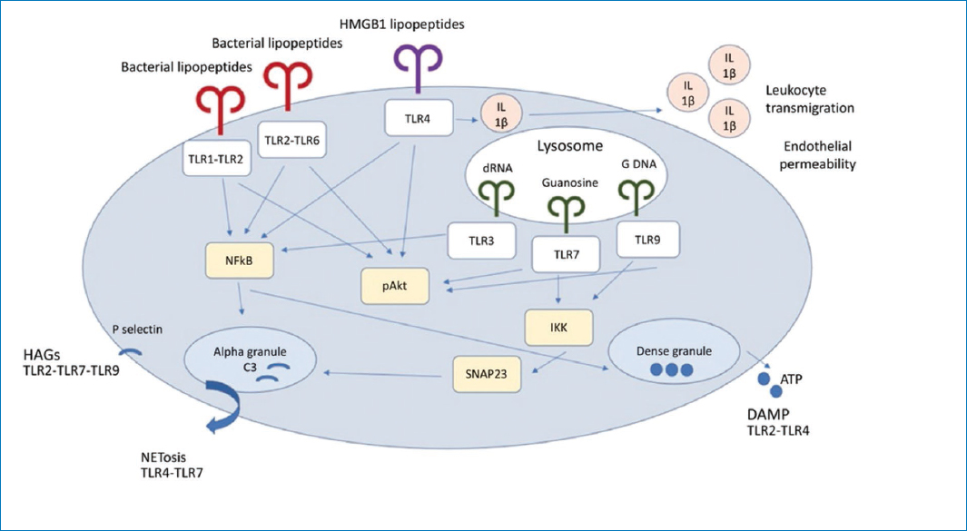
Figure 1. Activation of platelet TLRs. It induces rapid release or surface expression of proteins or molecules stored in platelet granules. Surface expression of P-selectin (or CD40L, in some cases) leads to interaction, in the form of heterotypic aggregates (HAG) with neutrophils and monocytes; HAGs are important for leukocyte activation and tissue transmigration. Platelets can also mediate (TLR7-complement C3 axis) or accelerate (TLR4) the NETosis process through their TLRs, either by direct or indirect interaction with neutrophils.
Platelets and TLR receptors
Surface TLRs (TLR2, TLR4 and TLR5) recognize pathogen surface components, while endosomal TLRs (TLR3, TLR7, TLR8 and TLR9) recognize pathogen nucleic acids. TLR10 is a surface TLR, but, unlike other TLRs, it suppresses inflammatory signaling in primary immune cells1 (Fig. 2).

Figure 2. Platelets and TLR receptors (bacterial capture by platelets).
The ability of platelets to generate and release IL-1b as a function of TLR4 suggests that, in situations of infection, platelets regulate endothelial barrier integrity, allowing other immune cells to enter the tissue and contain, confine and initiate responses to pathogens1.
Nucleotide-binding oligomerization domain (NOD) receptors
NOD 1 and 2 are also pattern recognition receptors, but unlike the TLR family, they are expressed in the cytoplasm. NOD receptors detect peptidoglycan components derived from the bacterial cell wall and provide defense against pathogen invasion rather than initial detection. NOD1 is activated as a result of g-d-glutamyl-meso-diaminopimelic acid (iE-DAP), while NOD2 detects the shorter fragment, muramyl dipeptide (MDP). In this sense, NOD1 responds specifically to gram-negative bacteria and some gram-positive bacteria, while NOD2 detects the cytoplasmic presence of any bacterium1.
Lectin receptors
C-type lectin receptors (CLR) are expressed on the cell surface and participate in immune responses to pathogens. CLRs recognize carbohydrate residues on the pathogen surface and can interact with TLRs on dendritic cells. Of the various CLRs described, platelets express functional DC-SIGN (CD209) and CLEC-2 (C-type lectin-like receptor 2). DC-SIGN can interact with pathogens by recognizing high levels of mannose and fucose on their surface. Platelets can also bind to HIV-1 particles using DC-SIGN, although CLEC-2 appears to be the predominant CLR involved in HIV-1 binding. Podoplanin is the main binding partner of CLEC-2, expressed on lymphatic endothelial cells and also on inflammatory macrophages. CLEC-2 activation leads to DC migration to lymph nodes and the T cell zone to initiate an immune response1.
Platelet-complement activation system
The complement system is activated directly in the circulation and consists of more than 30 proteins located in plasma. The ultimate goal of these two systems is the recognition of pathogens and the induction of defense responses that ultimately lead to the destruction and elimination of pathogens1.
Platelets can activate and be activated by the complement system. Platelets contain complement C3, C4a, C1 inhibitor (C1-I) and complement factor H in their a granules, and can release complement factor H and C1 inhibitor as a function of thrombin or collagen (C1-I) stimulation. C1 inhibitor inhibits activation of the complement cascade by interacting with activated C1 components, which are relevant to the classical pathway of activation. C1 also inhibits coagulation factors XIIa and FXIa, providing cross-regulation between coagulation and the complement cascade. The main function of complement factor H is to prevent spontaneous activation of the alternative pathway by mediating the dissociation of C3-convertase components that inhibit C3 hydrolysis and enhancing C3b cleavage, thereby inhibiting downstream activation of the lytic pathway. Interestingly, coagulation factor XIa can cleave complement factor H and, consequently, remove the brake on C3 activation and cascade stimulation. Complement regulation by FXIa provides another link of cross-regulation between the coagulation and complement pathways. Platelets can also activate the classical pathway of the complement cascade on their surface, which links innate and adaptive responses. The classical pathway of complement activation on the platelet surface is supported by the deposition of complement components C1q and C4d and the consequent generation of C4d and C3a1 (Fig. 3).

Figure 3. Platelets contain various molecules that initiate (C3) and amplify (Factor H, chondroitin sulfate [CS]) or inhibit (C1-i) the complement cascade. The alternative pathway is activated by spontaneous hydrolysis of complement C3 that is stabilized by pathogen surfaces and properdin and is amplified with complement factors B and D, or is inhibited with complement factors H and I. P-selectin on platelets can bind and activate C3b, it also supports activation of the complement system during the innate immune response. The classical pathway is initiated by IgM and IgG antibody complexes, as well as apoptotic surfaces.
Platelets and adaptive immune response
T cells are broadly classified as CD8+ cytolytic T cells or CD4 helper T cells. Because platelets are important sources of plasma cytokines and chemokines, platelets can play a role in mediating helper T cell differentiation.
Platelets interact directly with T cells and regulate the differentiation of antigen-presenting cells. Platelet-derived mediators, such as platelet factor 4, serotonin (5HT) and TGFb or transforming growth factor beta, influence helper T cell differentiation1.
Platelet extracellular vesicles are not always proinflammatory and may also have immunoregulatory potential. For example, they can provide 12-lipoxygenase to mast cells, which enhances lipoxin A4 production, a stimulator of inflammation resolution. Additionally, extracellular vesicles shed by stored platelets can polarize macrophages to an anti-inflammatory state. This effect may result from depletion of complement proteins (C1q, CFH, C3d). Platelet extracellular vesicles also regulate adaptive immunity: they can induce anti-inflammatory signaling in plasmacytoid dendritic cells and inhibit the differentiation of regulatory T cells into proinflammatory cells through a mechanism involving P-selectin1.
Platelets and NETosis
Platelets also mediate or shorten the time of NETosis. NETosis is a process that results in the release of DNA from neutrophils in response to the presence of pathogens or inflammatory stimuli. One of the most fascinating characteristics of neutrophils is their ability to release neutrophil extracellular traps (NET), web-like chromatin structures, whose main function is to entangle pathogens (specifically gram-negative bacteria) and eliminate them from the circulation. The release of neutrophil DNA together with platelets is important for capturing and eliminating bacteria or viral particles from the circulation. Neutrophil DNA is coated with positively charged histones that are prothrombotic. While beneficial for pathogen responses, this process, when dysregulated, is a double-edged sword and can contribute to thrombotic events and unstable coronary syndrome. NETs are potent inducers of thrombus formation, serving as a scaffold for platelet binding and activation and coagulation factors. As noted above, platelets, through their TLR4 receptor, can shorten NETosis time from hours to minutes. Platelets, through TLR7, mediate the level of NETosis by releasing complement C3 and further control their levels by elevating GM-CSF1.
Interactions of platelets with neutrophils, monocytes and macrophages
Platelet-neutrophil interactions are crucial in host defense and hemostasis. The number of NETs or platelet-neutrophil complexes (PNC) in the blood has been used as a marker of disease severity.
Ongoing systemic inflammation in immunological diseases may result in increased platelet-neutrophil interactions and a “vicious circle” of thromboinflammatory responses.
Platelets are equipped with receptors that allow them to detect vessel damage, inflammation or infection to become activated. Once activated, platelets can recruit leukocytes and interact with them, including monocytes and macrophages, stimulating mutual activation and cytokine release. Following activation, P-selectin (CD62P) is exposed on the platelet surface, where it can bind to its counter-receptor on myeloid cells PSGL1 (P-selectin glycoprotein ligand). This first association increases the expression of membrane-activated complex 1 (Mac-1) (CD11b/CD18 integrin aMb2) on the monocyte surface, which further supports their interactions with platelets. Upon activation, platelets adhere to monocytes through bridging molecules such as fibrinogen and thrombospondin. In addition to direct interactions, platelet-derived chemokines influence monocyte recruitment, adhesion and endothelial behavior1 (Fig. 4).

Figure 4. Activated platelet promoters of cytokines and chemokines to neutrophil.
The innate immune receptors of platelets are activated by immune complexes and bacterial-derived PAMPs, such as lipopolysaccharides, mediating immunopathogenic responses in sepsis. Platelet-derived serotonin, platelet-dependent NET secretion from glandular tissues and CLEC2 activation express podoplanin that play a fundamental role in tissue damage and shock in sepsis1.
Conclusion
Platelets are dynamic and crucial cells for prothrombotic and proinflammatory processes in different conditions, in addition to this they are equipped to participate in both innate and adaptive immune responses. As infections progress, platelets are also capable of responding to elevated levels of circulating antibody and pathogen components and sending signals to cells of the adaptive immune branch.
Funding
The authors declare that they have not received funding for this study.
Conflicts of interests
The authors declare no conficts of interests.
Ethical considerations
Protection of human subjects and animals. The authors declare that no experiments on humans or animals were performed for this research.
Confidentiality, informed consent, and ethical approval. This study does not involve personal patient data, medical records, or biological samples, and does not require ethical approval. SAGER guidelines do not apply.
Declaration on the use of artificial intelligence. The authors declare that no generative artificial intelligence was used in the writing or creation of the content of this manuscript.
