
Introduction
To maintain adequate organ perfusion, monitoring and control of effective mean arterial pressure (MAP) is necessary, which requires that the cardiovascular system be in proper functioning; however, in various clinical scenarios, whether originating from sepsis, myocardial dysfunction, severe trauma, hemorrhage, various shock states, etc., cardiovascular dysfunction may be generated.
Organ hypoperfusion can trigger severe repercussions such as ischemia and infarction, which is why it is necessary to maintain a minimum MAP of 60 mmHg, as recommended in several clinical guidelines; if this decreases for prolonged periods, it can cause irreversible damage (Fig. 1)1.
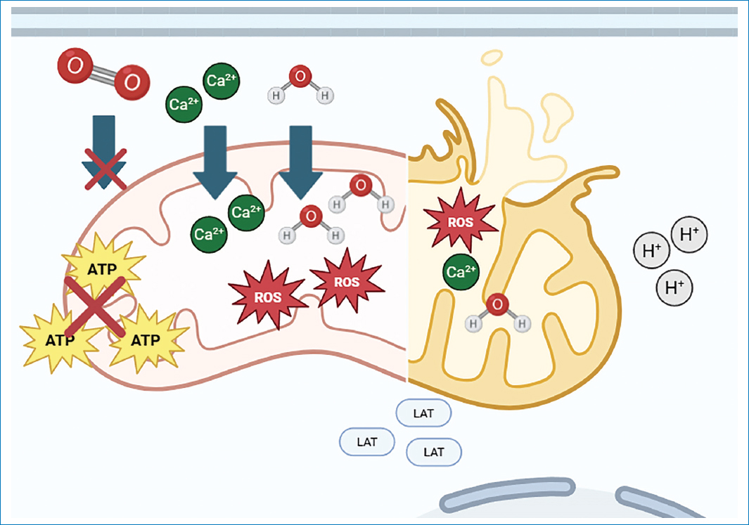
Figure 1. Schematic representation of the pathophysiology of ischemia-induced mitochondrial damage. The diagram illustrates the cascade of deleterious cellular events following O2 deprivation. In the initial phase (left half), hypoxia halts the electron transport chain, resulting in an abrupt drop in ATP synthesis (crossed-out stars). This bioenergetic failure incapacitates transmembrane ionic pumps, provoking a massive influx of Ca2+ and H2O toward the mitochondrial matrix, initiating edema. With damage progression (right half), severe architectural disruption is observed with loss of mitochondrial cristae and overproduction of ROS (red stars), which exacerbate damage to lipid membranes. At the cytosolic level, metabolism shifts toward anaerobic glycolysis, evidenced by accumulation of LAT and H+, which conditions a state of intracellular acidosis that contributes to irreversible cell death. ATP: adenosine triphosphate; Ca2+: calcium; H2O: water; O2: oxygen; LAT: lactate; H+: protons; ROS: reactive oxygen species.
Definitions
Blood pressure
It is defined as “the force exerted by blood against the walls of the arteries during its circulation propelled by the heart.” This pressure is the result of an interaction between three important variables: cardiac output, blood volume, and systemic vascular resistance; their adequate relationship guarantees good perfusion of tissues and vital organs2.
It is expressed through two components: systolic blood pressure – the maximum pressure in the arteries during ventricular contraction – and diastolic blood pressure – the minimum pressure during cardiac relaxation. Both determine MAP.
MAP
It is defined as the average pressure in the arteries during a complete cardiac cycle that reflects an effective perfusion pressure of vital organs. It encompasses the cardiac cycle, systole, and diastole and is in turn influenced by cardiac output and systemic vascular resistance.
One of the most commonly used formulas is MAP = SBP + 2(DBP)/3. This is why it is important to recognize several terms, as maintaining a MAP below normal values can be related to various factors such as hypotension (vasodilation), whether due to sepsis or anesthesia, low cardiac output, hypovolemia, etc.3,4.
Intra-abdominal pressure (IAP)
It is the pressure existing within the abdominal cavity, resulting from the balance between abdominal content (viscera, fluids, gas) and the distensibility of the abdominal wall2. It is measured with the patient in supine position at the end of expiration with muscle relaxation; values are found in healthy adults at 2-7 mmHg, sick adults at 5-7 mmHg, in obesity, and pregnancy at 10-15 mmHg4. The abdominal cavity is a closed compartment that is limited by the diaphragm (superior), vertebral column and dorsal muscles (posterior), abdominal wall (anterior), and pelvis, and sexual and urinary organs (inferior). When a person has a pathology or physiological change that influences the change in these compartments, IAP can be modified, either its increase or decrease in distensibility (example: abdominal tumors, pregnancy, obesity, burns, surgical procedure, etc.). IAP is not constant, as it is modified by various situations such as sustained respirations, cough, certain maneuvers such as Valsalva, mechanical ventilation, and abdominal contraction, etc.5 (Table 1 and Fig. 2).
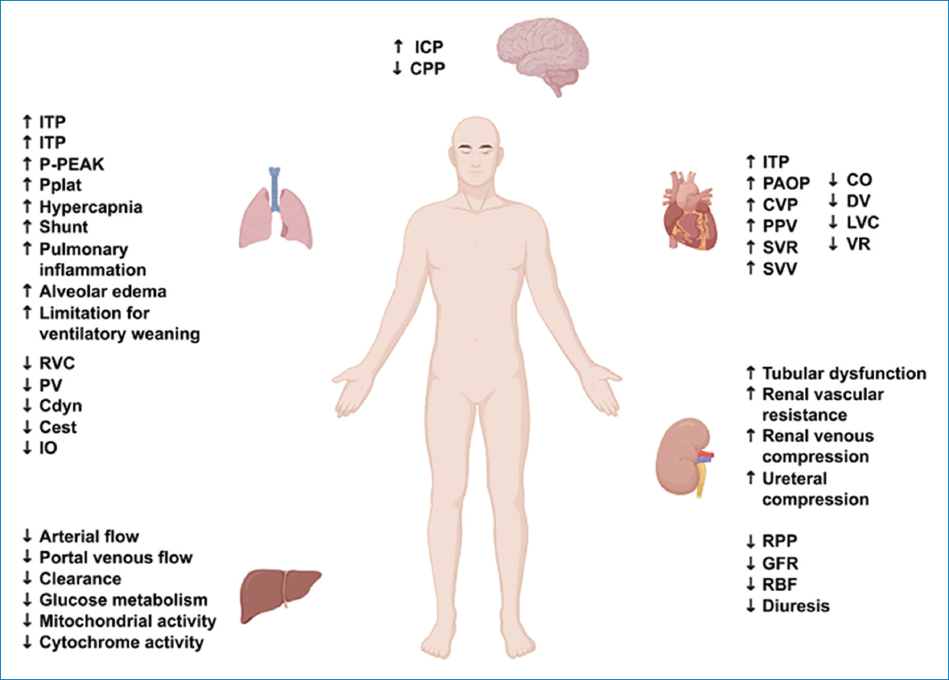
Figure 2. Pathophysiological changes of IAH and ACS. ICP: intracranial pressure; CPP: cerebral perfusion pressure; ITP: intrathoracic pressure; PP: pleural pressure; P-PEAK: peak airway pressure; Pplat: plateau pressure; RVC: residual vital capacity; PV: pulmonary volume; Cdyn: dynamic compliance; Cest: static compliance; OI: oxygenation index; PAOP: pulmonary artery occlusion pressure; CVP: central venous pressure; PPV: pulse pressure variation; SVR: systemic vascular resistance; SVV: stroke volume variation; CO: cardiac output; DV: diastolic volume; LVC: left ventricular compliance; VR: venous return; RPP: renal perfusion pressure; GFR: glomerular filtration rate; RBF: renal blood flow.
Table 1. Systemic affection secondary to hypoperfusion due to decreased mean arterial pressure
| System | Function | Mechanism of alteration | Injury |
|---|---|---|---|
| Hemodynamic | Global tissue perfusion | ↓ Pressure gradient | Systemic hypoperfusion Brain: ↓ CPP → confusion/ischemia Kidney: ↓ GFR → oliguria/AKI Liver: ↓ flow → ↑ transaminases Heart: ↓ coronary perfusion → ischemia |
| Oxygenation and transport | O2 delivery (DO2) | ↓ Blood flow | Cellular hypoxia; lung: V/Q imbalance → hypoxemia |
| Metabolic | Energy production | Shift to anaerobic glycolysis; ↓ O2 + acidosis | ↑ Lactate; ↓ ATP |
| Ionic and cellular | Na⁺/K⁺and Ca2⁺pumps | ↓ ATP → pump failure | Cellular edema + ↑ Ca2⁺ |
| Oxidative | Redox balance | ↑ ROS (especially in reperfusion) | Damage to lipids, proteins, and DNA; Microcirculation: stasis → dysfunction |
| Structural (membranes) | Cellular and organelle integrity | Phospholipases+Ca2⁺ + edema | Membrane rupture; coagulation: endothelial damage → coagulopathy |
|
CPP: cerebral perfusion pressure; GFR: glomerular filtration rate; AKI: acute kidney injury; DO2: oxygen delivery; V/Q: ventilation/perfusion ratio; ROS: reactive oxygen species; ATP: adenosine triphosphate; ↑ Increases; ↓ Decreases; → Produces; Ca2+: calcium; O2: oxygen; Na+: sodium ion; K+: potassium ion. |
|||
Abdominal perfusion pressure (APP)
It is defined as the relationship that exists between MAP and IAP. It has been shown to be a much more precise marker of survival than guiding resuscitation strategies solely with MAP or IAP. It is described in the literature that APP values below 60 mmHg showed significantly higher mortality in the short and long term in correlation with values above this threshold5,6. Therefore, active measurement of APP and its use as an additional guide in the vasopressor management of patients can favor better clinical outcomes for the group of hemodynamically unstable patients, thus favoring safer withdrawal of vasopressors compared to the classical measurement of MAP, especially to avoid abdominal hypoperfusion in scenarios where IAP is increased.7
APP is defined as:
APP = MAP-IAP
In anesthesia, it is of important relevance due to the effect on patient ventilation when subjected to general anesthesia, from increased peak pressure and decreased pulmonary compliance; hemodynamically, it decreases venous return and cardiac output; in renal perfusion, there is intraoperative oliguria; in laparoscopic surgeries, IAP can rise to levels between 12 and 15 mmHg, producing even more hemodynamic and ventilatory changes, leading to decreased APP and the need for vasopressor medication8.
The variability between APP values is based on the two components from which the calculation is obtained, which are MAP and IAP; it is important to remember that normal IAP is between 5 and 7 mmHg8,9 (Table 2).
Table 2. Normal and pathological values of intra-abdominal pressure
| Category | p | Context |
|---|---|---|
| Physiological (healthy adult) | ≤ 5 mmHg | Considered normal in adults |
| Range in critical patient | 5-7 mmHg | Expected values in critically ill patients |
| Elevated without pathological significance | 10-15 mmHg | May be observed without implying pathology (e.g., obese patients) |
| IAH | > 12 mmHg | Defined by elevation in 3 consecutive measurements (every 4-6 h) |
| ACS | > 20 mmHg | Sustained elevation+organ dysfunction or failure |
|
IAH: intra-abdominal hypertension; ACS: abdominal compartment syndrome. |
||
Role of APP and the use of vasopressors in the critical patient
There is a great dilemma with the use of vasopressors, especially in two aspects: first, the time during which it is necessary to maintain the patient’s clinical stability, and second, the correct moment and method for de-escalation and withdrawal.
Since their use is associated with various adverse effects such as tachycardia, excessive vasoconstriction (favoring tissue ischemia scenarios), cardiac arrhythmias, metabolic and even immunological alterations; therefore, perfusion and de-escalation strategies have been devised in various protocols seeking to establish a goal at which their withdrawal can be considered safe, and among these, the use of MAP usually has a central role6–11 (Fig. 3).

Figure 3. Mean arterial pressure-based management algorithm.
Typically, the use of MAP could be considered an efficient standard within the majority of clinical scenarios; however, given the importance of safe environments for the clinical recovery of patients and the diversity of pathologies that require the use of vasopressor medication, scenarios may be found in which a MAP does not translate into adequate tissue perfusion; and it is in these scenarios where concepts such as APP gain value10–15.
The importance of measuring APP is based on the high incidence of intra-abdominal hypertension (IAH) in critically ill hospitalized patients. In a study conducted by Vidal et al., it was reported that patients in the intensive care unit presented an incidence of this entity in 64% at any time during their stay, also reporting that the main risk factors were mechanical ventilation, respiratory distress, and exhaustive fluid resuscitation16. As abdominal hypertension is a frequent entity in critically ill patients or in the intensive care unit, in addition to being an early sign of multiorgan failure and a marker of survival, its use in the titration of vasopressor medication as part of APP is justified, especially to avoid relying solely on MAP as a resuscitation target, since in scenarios with abdominal hypertension, this could generate errors in resuscitation objectives17,18 (Table 3).
Table 3. Recommended treatment for intra-abdominal pressure
| IAP level | Definition | Anesthetic objective | Specific treatment (escalated) | Anesthetic considerations |
|---|---|---|---|---|
| Normal (0-5 mmHg) | Physiological | Maintain perfusion (APP > 60 mmHg) | Surveillance | Avoid fluid overload |
| IAH Grade I (12-15 mmHg) | Mild hypertension | Prevent progression | Optimize analgesia and sedation Avoid excessive PEEP Neutral position (avoid Trendelenburg) | Adjust ventilation (↓ Peak pressure) |
| IAH Grade II (16-20 mmHg) | Moderate | Decrease intra-abdominal pressure | Deep sedation Neuromuscular blockade Gastric/rectal decompression Negative fluid balance | ↓ Pulmonary compliance, ↑ Plateau pressure |
| IAH Grade III (21-25 mmHg) | Severe | Restore organ perfusion | Diuretics/ultrafiltration, paracentesis if ascites Percutaneous drainage Avoid excessive crystalloids | Risk of ↓ venous return, hypotension |
| ACS (> 20 mmHg + organ dysfunction) | Abdominal compartment syndrome | Urgent decompression | All previous measures Decompressive laparotomy (Gold standard) Open abdomen (VAC) | Critical induction: high risk of hemodynamic collapse |
|
IAP: intra-abdominal pressure; APP: abdominal perfusion pressure; IAH: intra-abdominal hypertension; ACS: abdominal compartment syndrome; PEEP: positive end-expiratory pressure; VAC. vacuum-assisted closure. |
||||
Increased IAP values negatively impact various systems, reducing venous return, increasing inferior vena cava pressure, decreasing cardiac output, favoring oliguria and acute kidney injury, elevating the diaphragm, reducing pulmonary capacity, favoring hypoxia, and even being able to increase intracranial pressure by hindering jugular venous drainage3,4,6,10,16,19.
Increases in IAP are framed not only in the absolute values expressed in millimeters of mercury but also in a fine relationship between abdominal content and the abdomen’s capacity to stretch (abdominal compliance). The measurement of IAP through invasive elements commonly used in patients, such as bladder catheter, can provide reliable data for the calculation of APP9 (Fig. 4).
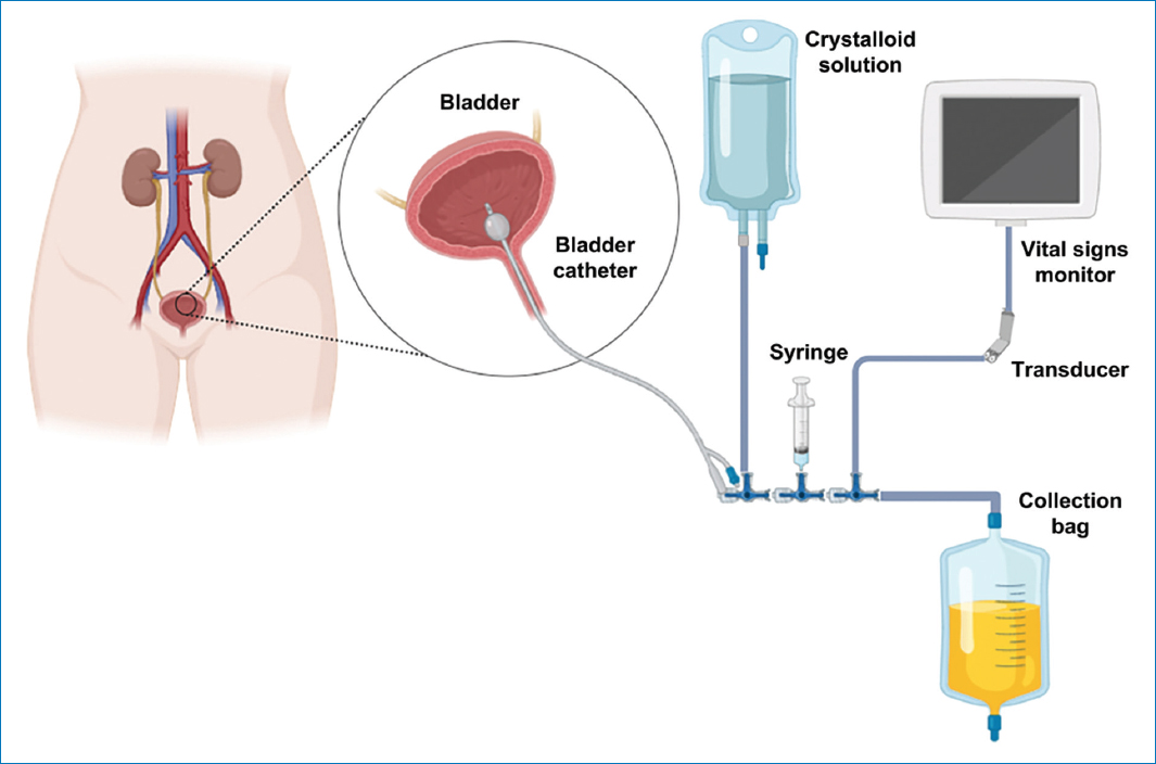
Figure 4. Diagram of intra-abdominal pressure measurement through bladder.
Optimization of APP > 60 mmHg, as well as its use as a marker in the management with vasopressors and fluid resuscitation therapy, allows better control of intestinal edema, compression of renal vessels, and tissue ischemia, reducing the risk of complications and clinical deterioration associated with overly aggressive resuscitations20.
Use of APP in the withdrawal of vasopressors
The optimal sequence for vasopressor withdrawal in vasodilatory shock has been explained based on the pathophysiological model proposed by Landry et al., which distinguishes between two partially independent mechanisms of vasodilation: the catecholamine-sensitive pathway and a state of relative vasopressin deficiency21,22.
In septic shock, endogenous vasopressin concentrations initially rise but subsequently decrease due to depletion of neurohypophyseal stores and alteration of its baroreceptor-mediated release, generating a state of relative vasopressin deficiency22. This phenomenon contributes to persistent vasodilation that does not respond completely to catecholamines. In this context, exogenous vasopressin administration acts as hormonal replacement therapy, restoring vascular tone through V1 receptors, which remain relatively preserved during shock21,22. On the other hand, the catecholamine response, mediated primarily by α-adrenergic receptors, tends to recover earlier as the inflammatory state improves, despite the desensitization observed in initial phases21 (Fig. 5).
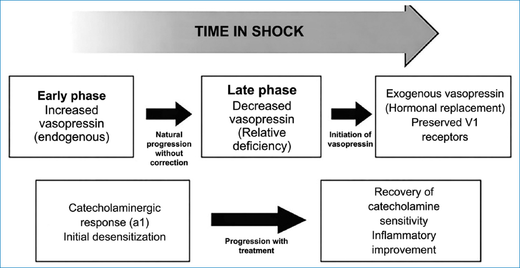
Figure 5. Shock dynamics.
Based on this difference in the recovery of vasopressor systems, early discontinuation of vasopressin may unmask persistent deficiency and precipitate hypotension, while norepinephrine can be withdrawn more safely in the initial stages.
This physiological foundation is supported by clinical evidence. Post hoc analyses of the VASST study and subsequent observational studies have demonstrated a higher incidence of hypotension when vasopressin is discontinued before norepinephrine23. Therefore, several experts recommend a strategy in which norepinephrine is reduced first, maintaining low doses of vasopressin until hemodynamic stability is ensured.
From a clinical perspective, the interaction between vasopressor withdrawal and IAP may modify patient management. Consider a patient recovering from septic shock who remains on low doses of norepinephrine and vasopressin. In a scenario with normal IAP, progressive reduction of norepinephrine while maintaining vasopressin is usually well tolerated, as improvement of the inflammatory state allows recovery of catecholamine sensitivity. Once norepinephrine is discontinued and hemodynamic stability is maintained, vasopressin can be withdrawn cautiously21,22,24,25 (Fig. 6).
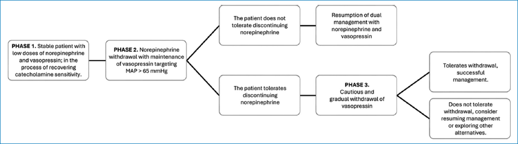
Figure 6. Sequential strategy for vasopressor support withdrawal in the critical patient with normal intra-abdominal pressure.
In patients with increased IAP, the situation may differ significantly. Increased IAP reduces APP and may compromise venous return, contributing to persistent circulatory instability despite apparent resolution of the inflammatory state26,27. In this context, vasopressin may play a particularly relevant role in maintaining vascular tone through non-adrenergic mechanisms. Therefore, early discontinuation of vasopressin in these patients may trigger hypotension, even when norepinephrine requirements are low28–30. Therefore, persistent mechanical factors, such as IAH, must be considered when planning vasopressor withdrawal10,21–25 (Fig. 7).
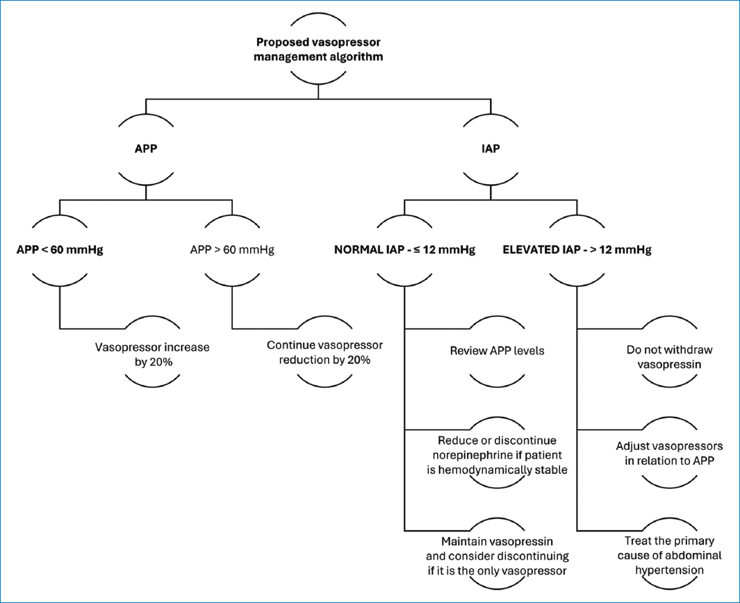
Figure 7. Proposed algorithm for management based on APP and IAP measurement. APP: abdominal perfusion pressure; IAP: intra-abdominal pressure.
Clinical implications and limitations
APP has been proposed as a physiological parameter that reflects the effective perfusion gradient of abdominal organs in critically ill patients. This concept is analogous to that of cerebral perfusion pressure, which integrates systemic arterial pressure and intracranial pressure to estimate the effective driving force of cerebral blood flow. Similarly, APP attempts to consider the mechanical effects of increased IAP on the perfusion of abdominal organs, particularly in patients with IAH or abdominal compartment syndrome (ACS).
The incidence, risk factors, and outcomes of intra-abdominal study: intra-abdominal pressure: risk factors and outcomes, a multicenter prospective investigation, reported that IAH occurs in about half of patients admitted to intensive care units and that its severity independently predicts mortality. Because APP integrates both systemic arterial pressure and IAP, it provides a physiologically more complete assessment of abdominal perfusion than either of these variables separately.
APP has limitations as a perfusion marker; it continues to be an indirect macrohemodynamic parameter and should not be considered a direct measurement of tissue perfusion. Tissue perfusion depends not only on perfusion pressure but also on microcirculatory function, capillary density, endothelial integrity, and cellular oxygen utilization. In conditions such as septic shock, significant microcirculatory dysfunction may persist despite apparently adequate perfusion pressures. In addition, most of the clinical evidence supporting the use of APP comes from observational studies rather than randomized clinical trials. Although associations between low APP values and organ dysfunction are consistent, it is not yet clear whether directing resuscitation specifically toward APP goals improves clinical outcomes compared to traditional MAP-guided strategies. In accordance with these limitations, the guidelines published in 2013 by the World Society of the ACS concluded that available evidence was insufficient to recommend routine monitoring of APP as a resuscitation target.
The relationship between APP and vasopressor therapy is particularly relevant during the management of septic or distributive shock. Vasopressors increase MAP and, consequently, can improve APP when IAP remains constant. In patients with elevated IAP, vasopressors can partially compensate for the reduction in APP by increasing arterial driving pressure.
Vasopressor therapy represents a double-edged strategy in this context. Although increased MAP may improve APP, excessive vasoconstriction may simultaneously deteriorate regional microcirculatory blood flow. Experimental and clinical studies suggest that high doses of catecholamines can reduce mesenteric perfusion and contribute to the development of intestinal ischemia in vulnerable patients. Therefore, increasing vasopressor doses with the objective of maintaining APP, without addressing the elevation of IAP, may not restore effective tissue perfusion.
This has implications for vasopressor withdrawal. During the shock stabilization phase, progressive reduction of vasopressor support is usually attempted as organ perfusion improves. However, in patients with persistent IAH, reductions in MAP may cause disproportionate decreases in APP and subsequent deterioration of renal or splanchnic perfusion. Recognizing this interaction may help interpret apparent vasopressor dependence. Instead of solely increasing vasopressor doses, management should also be oriented toward identifying and treating reversible causes of elevated IAP, such as excessive fluid accumulation, gastrointestinal distension, abdominal wall rigidity, or the presence of intra-abdominal collections. Strategies to reduce IAP include gastric and colonic decompression, optimization of sedation and analgesia, avoiding excessive fluid administration, promoting diuresis or fluid restriction when appropriate, and drainage of ascites or intra-abdominal collections. In cases of ACS with organ dysfunction, decompressive laparotomy may be necessary. By addressing both components of the APP equation – arterial pressure and IAP – it is possible to facilitate safer vasopressor withdrawal and simultaneously preserve abdominal organ perfusion.
Conclusion
APP should be considered a useful complementary marker in the hemodynamic evaluation of patients with suspected or diagnosed IAH rather than a universal resuscitation target. Despite its solid pathophysiological basis, the role of APP as a therapeutic target in the critical patient is not yet completely defined. Most of the available evidence comes from observational studies or cohorts with limited sample sizes, and there is currently a shortage of high-quality randomized clinical trials evaluating APP-guided resuscitation strategies.
Funding
The authors declare that they have not received funding.
Conflicts of interest
The authors declare no conflicts of interest.
Ethical considerations
Protection of human subjects and animals. The authors declare that no experiments on humans or animals were performed for this research.
Confidentiality, informed consent, and ethical approval. This study does not involve personal patient data, medical records, or biological samples, and does not require ethical approval. SAGER guidelines do not apply.
Declaration on the use of artificial intelligence. The authors declare that no generative artificial intelligence was used in the writing or creation of the content of this manuscript.
