Introduction
Parkinson’s disease (PD) is a central clinical motor syndrome accompanied by neurodegeneration of the substantia nigra pars compacta (SNc) and the deposition of synuclein.1 Its prevalence is estimated at 1% among individuals over 60 years of age.2 For symptomatic treatment, systemic administration of L-3,4-dihydroxyphenylalanine (L-Dopa) is commonly used owing to its efficacy. Prolonged L-Dopa treatment is associated with L-Dopa-induced dyskinesias (LIDs).3 LIDs are abnormal involuntary movements (AIMs) with a prevalence of 34% in patients with PD,4 an incidence of 40% after 4 years of L-Dopa treatment,5 and up to 80% after 10 years.6
In the basal ganglia, LIDs are associated with cellular, molecular, and functional alterations. These include a redistribution of dopaminergic D1 receptors (D1Rs), potentiation of intracellular signaling, and dysregulated gene expression in neurons expressing D1Rs, which contributes to pre- and post-synaptic changes in dopaminergic transmission, such as increased receptor sensitivity, abnormal synaptic activity, and elevated neurotransmitter levels.7 This results in a neurotransmitter imbalance involving gamma-aminobutyric acid (GABA) and glutamate in the corticostriatal pathway, with an increase in the release of GABA and glutamate in the striatum of parkinsonian rats with LIDs.8 Striatal glutamate is released by cortical or thalamic glutamatergic axons.9 The increase in glutamate levels in the corticostriatal and thalamocortical pathways is characteristic of hyperkinetic disorders such as LIDs.
The striatum receives hypothalamic histaminergic innervation and exhibits high expression of histamine H3 receptors (H3Rs).10 H3Rs are co-expressed with dopaminergic receptors in GABAergic medium spiny neurons (MSNs). Specifically, H3Rs are found together with D1Rs in the MSNs that give rise to the direct striatonigral pathway and with D2 receptors in those that form the indirect striatopallidal pathway.11 Multiple studies support a functional interaction between D1Rs and H3Rs at the membrane of MSNs of the direct pathway.12 Presynaptically, H3R activation by the agonist immepip reduces the D1R-dependent component associated with depolarization-evoked (3H)-GABA release in striatal and rat substantia nigra pars reticulate slices.13 Post-synaptically, H3R stimulation attenuates D1R-induced cAMP signaling, whereas D1R activity appears to potentiate H3R-mediated activation of mitogen-activated protein kinases, highlighting a bidirectional modulatory relationship between these receptor systems.14
Previous studies have demonstrated that acute systemic administration of the selective H3R agonist immepip (1-10 mg/kg, s.c.) does not reduce LIDs in rats with 6-hydroxydopamine (6-OHDA) lesions in the SNc.8,15 According to the classical dyskinesia circuit, overactivation of thalamocortical and corticostriatal glutamatergic afferents drives hyperactivity of the direct striatonigral pathway, thereby contributing to LIDs.16 However, drugs that reduce glutamatergic transmission, such as amantadine, MTEP, and MK-801, have shown antidyskinetic properties.17 In this context, H3R activation could exert beneficial effects by counteracting glutamatergic overactivity and reducing dyskinesias, potentially through functional interactions with D1Rs, which facilitate GABA release in the striatum and modulate excitatory signaling in MSNs.18
In the present study, we examined the impact of chronic H3R activation on dyskinesias induced by chronic L-Dopa administration in 6-OHDA-lesioned rats.
Material and methods
Male Wistar rats weighing 280-310 g, obtained from the Laboratory Animal Production and Experimentation Unit (UPEAL, Cinvestav), were used. Animals were acclimatized to standard laboratory conditions and maintained under a 12-h light/dark cycle. All experimental procedures were performed in accordance with the guidelines of the Guide for the Care and Use of Laboratory Animals19 and were approved by the Animal Care Committee of the Instituto Nacional de Rehabilitación (protocol 50/23). To ensure the ethical use of animals, the minimum number required was employed in accordance with the bioethical and statistical recommendations established by Festing.20
6-OHDA lesion
Rats were anesthetized with a ketamine-xylazine combination (80/10 mg/kg, i.p.) and placed in a stereotaxic apparatus (Stoelting Corp., Wood Dale, IL, USA). A midline scalp incision was made, the skull was exposed, and a craniotomy of approximately 1 mm was performed at the following stereotaxic coordinates: anteroposterior −5.0 mm from bregma, lateral (L) −2.0 mm from the midline, and vertical (V) −7.2 mm from the dura mater, according to Paxinos and Watson.21 To induce a unilateral SNc lesion, 6-OHDA (8 μg/μL in saline containing 0.1% ascorbic acid) was injected at a rate of 0.4 μL/min over 5 min using a microinjection pump (CMA/400, CMA Microdialysis, Kista, Sweden). The needle (30 gauge) was left in place for an additional 5 min after the injection to allow adequate diffusion of the neurotoxin and prevent backflow. Three weeks later, the efficacy of the dopaminergic lesion was assessed by the apomorphine-induced rotational test (0.5 mg/kg, s.c.); only animals displaying six or more contralateral rotations per minute were selected for the study.
Experimental design
Forty hemiparkinsonian rats were randomly assigned to four groups (10 animals per group):
- − The vehicle group (6-OHDA; vehicle) received daily intraperitoneal (i.p.) injections of saline for 14 consecutive days
- − The L-Dopa-only group (6-OHDA; L-Dopa), in which each rat was pretreated with benserazide (15 mg/kg, i.p.) followed 20 minutes later by L-Dopa (6.25 mg/kg, i.p.) for 21 days
- − The chronic immepip group (6-OHDA; L-Dopa+immepip), which received L-Dopa in combination with immepip (1 mg/kg, i.p.) for 14 days
- − The immepip withdrawal group (6-OHDA; immepip withdrawal) was treated daily with L-Dopa plus immepip for 14 days; on the following day, immepip was discontinued, and animals continued receiving L-Dopa alone for an additional 7 days until day 21 post-treatment. In all groups receiving combination treatment, L-Dopa administration was consistently preceded by the corresponding peripheral decarboxylase inhibitor.
Behavioral analysis
AIMs were evaluated in all animals using a scale previously established and validated by Cenci et al.,22 which distinguishes four dyskinesia subtypes: axial, limb, orolingual, and locomotive. Each subtype was scored on a 0-4 scale according to the specific criteria of Cenci et al.22 0 = absent; 1 = occasional; 2 = frequent; 3 = continuously present but suppressible by external stimuli; and 4 = continuously present and not interrupted by sensory distraction. Evaluations were performed once daily, recording AIMs for 1 min every 20 min over a 120-min period, beginning 20 min after L-Dopa treatment. Thus, six observation periods per session were analyzed, and individual scores were summed, yielding a maximum possible score of 24 per AIM subtype. The total number of AIMs was tallied for each subtype. Locomotive AIMs were excluded from the final analysis owing to the ongoing debate regarding their interpretation.22 All sessions were video-recorded and behavioral evaluations were performed by an investigator blinded to the experimental conditions.
Statistical analysis
All data are presented as mean ± standard error of the mean, except for the results shown in figure 1A–C, which were reported as median ± interquartile range. For statistical comparisons of AIM scores, non-parametric tests were applied: the Kruskal-Wallis test followed by post hoc Mann-Whitney U or Dunn’s tests, depending on the context of the analysis. Total AIMs were analyzed by repeated-measures analysis of variance (ANOVA) with Bonferroni post hoc correction to evaluate differences among pharmacological treatment groups. For the immepip withdrawal group, changes in AIM scores over time were evaluated using the Wilcoxon signed-rank test, while total AIMs in this group were compared using Student’s t-test. A p-value below 0.05 was considered indicative of statistically significant differences.
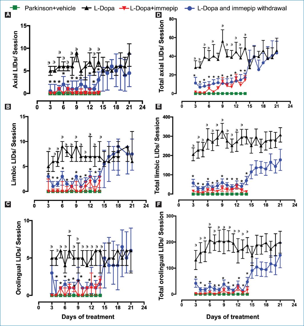
Figure 1. Effect of chronic administration of the H3 receptor agonist immepip (1 mg/kg, i.p.), and its withdrawal, on L-Dopa-induced dyskinesias (LIDs) (L-Dopa 6.25 mg/kg/benserazide 15 mg/kg, i.p.): A: axial LID scores B: limbic (limb) LID scores C: and orolingual LID scores D: and total axial LIDs E: limb LIDs F: and orolingual LIDs. The chronic immepip treatment group received a daily dose of L-Dopa + immepip (1 mg/kg, i.p.) for 14 days (red lines), and in the immepip withdrawal group, immepip was withdrawn starting from day 14 (blue line). Statistical analysis of abnormal involuntary movements (AIM) scores was performed using the non-parametric Kruskal-Wallis test, followed by Dunn’ multiple comparisons test. Total AIM analysis was performed by repeated-measures analysis of variance, followed by Bonferroni post hoc test to compare means across pharmacological treatment groups (10 animals per group).3 p < 0.01, compared with the vehicle group; *p < 0.01, compared with the L-Dopa-treated group.
Results
In 6-OHDA-lesioned rats, daily administration of L-Dopa for 14 days at a dose of 6.25 mg/kg consistently induced dyskinesias in all animals, with scores ranging from 1 to 3 (Fig. 1A–F), reliably reproducing the LID model. Chronic immepip administration counteracted the dyskinetic effect of L-Dopa and led to infrequent-to-normal dyskinetic movements with scores of 0 and 1 in 75% of the rats (Fig. 1A–F). However, withdrawal of the agonist immepip from days 15 to 21 increased the incidence of dyskinetic movements by up to 143%, with scores returning to the 1-3 range, similar to the L-Dopa-only group. In contrast, no abnormal movements were observed in the vehicle group. These results were also reflected in the daily AIM scores: L-Dopa significantly increased total LIDs and axial, limb, and orolingual scores from day 2 onwards, while chronic immepip treatment consistently reduced these scores (Fig. 1A–F).
Discussion
In the present study, the beneficial effects of systemic administration of the H3R agonist immepip were reproduced through reductions in both total LIDs and scale scores.8,23 Administration of L-Dopa alone significantly increased total AIMs and their scores across all three subtypes: axial, limb, and orolingual. In contrast, chronic treatment with immepip (for 14 days) consistently reduced the expression of all AIM subtypes in the L-Dopa+immepip group (Fig. 1). This effect was reversed following immepip withdrawal, as observed in the immepip withdrawal group. It is important to note that the chronic immepip administration group showed stable values, consistent with our previous results.8,23 Indeed, immepip was administered for 14 days; however, its effect is reversible upon drug withdrawal, as observed during its discontinuation from days 15 to 21 (Fig. 1). In humans, the long-term antidyskinetic efficacy of other drugs, such as amantadine, has been widely recognized.24 A future long-term study comparing the effects of amantadine and immepip may be warranted to explore the efficacy of immepip, given the inherent limitations associated with the use of an animal model. Indeed, it has been found that repeated amantadine administration in animals produces a reduction of its antidyskinetic effect.25 Amantadine withdrawal leads to the reappearance of LIDs, which can reach a high intensity.26 In our studies,8,23 including the present one, the antidyskinetic effect of immepip disappears upon drug withdrawal; however, the intensity of AIMs does not exceed the values observed in the L-Dopa-only group.23
According to Bastide et al.,27 “priming” refers to abnormal neurochemical and functional changes within dopamine-depleted basal ganglia. In dyskinesia, this phenomenon arises owing to basal ganglia abnormalities triggered by the administration of dopaminergic agents such as L-Dopa.28 In the present study, we propose that this antidyskinetic phenomenon is triggered at the striatal level through the functional interaction between dopamine D1Rs and histamine H3Rs. However, further studies are needed to demonstrate this hypothesis at the molecular and functional levels.12
Conclusion
The antidyskinetic effect of the H3R agonist immepip is explained by the functional interaction between D1Rs and H3Rs, both of which are colocalized in the striatum, although we do not rule out a potential intrastriatal effect. Future studies are needed to examine the amino acid levels, such as GABA and glutamate, in the thalamocortical pathway, with the aim of determining whether the antidyskinetic effect is accompanied by a reduction in the activity of this pathway.
Funding
The authors declare that they received no funding for this study.
Conflicts of interest
The authors declare no conflicts of interest.
Ethical considerations
Protection of human subjects and animals. The authors declare that the procedures followed were in accordance with the ethical standards of the responsible committee on human experimentation and with the World Medical Association and the Declaration of Helsinki. The procedures were authorized by the Institutional Ethics Committee.
Confidentiality, informed consent, and ethical approval. This study does not involve personal patient data, medical records, or biological samples, and does not require ethical approval. SAGER guidelines do not apply.
Declaration on the use of artificial intelligence. The authors declare that no generative artificial intelligence was used in the writing or creation of the content of this manuscript.
