
Introduction
Fanconi anemia (FA) is a rare genetic disorder associated with profound genomic instability and defects in DNA repair pathways involving the repair of crosslinking between strands. FA occurs in approximately 1 in 200,000 newborns worldwide, with an estimated incidence of 4-7 cases/million live births in Europe1. Despite such a molecular deficiency confers vulnerability for a broad spectrum of hematological diseases and solid tumor, only 15% of Fanconi’s anemia patients develop malignancies. These are chiefly acute myeloid leukemia and myelodysplastic syndrome, as well as a range of other solid tumors and liver tumors. According to the Cancer Genome Atlas database, the prevalence of somatic mutations in Fanconi anemia complementation group A (FANCA) genes represents ∼3% for gastric cancer. FANCA, FANCB, FANCC, FANCE, FANCG, FANCL, and FANCM comprise the FANC family, all representing a variety of genes, making FA a heterogeneous genetic disease. Of these genes, FANCA is the most common and contains a 1455-amino acid nuclear protein and plays a role in a multiprotein complex necessary for DNA repair and chromosomal stability.
Non-synonymous genetic variations (non-synonymous single-nucleotide polymorphisms [nsSNPs]) affecting FANCA carry a specific interest as they modify the amino acid order of the protein and also alter the protein physicochemical and structural properties. These deleterious mutations lead to damage to the repair complex, causing genomic instability and vulnerability to malignant transformation2.
Similarly, the FANCI gene is involved in the FA repair pathway as a complementary pathway. These pathogenic variants in FANCI impair DNA crosslink repair, leading to progressive bone marrow failure where neoplasms arise from a higher susceptibility3.
The Fanconi anemia complementation Group D2 (FANCD2)-FANCI interaction through monoubiquitination plays an essential regulatory role in DNA repair, and the dysregulation of FANCD2-FANCI activates cellular stress pathways (ATR-CHK1) and augments alternative, error-prone repair mechanisms, which favors accelerated mutagenesis4.
The accumulation of somatic mutations and a change of cell cycle checkpoints promote neoplastic transformation in FA4.
Gastric cancer has been described as one of the frequent solid manifestations in patients with FA; it is characterized by early onset (i.e., 30-40 years) and high histological aggressiveness5.
Case reports6,7 demonstrate that even heterozygous variation in genes like FANCA and FANCD2 may dramatically increase predisposition to gastric adenocarcinoma, indicating that incomplete FA pathway activation will suffice for gastric carcinogenesis activation.
A case about 25-years-old male with FA and Gastric carcinoma is presented.
Case report
We report a 25-year-old male who was diagnosed at 14 years of age with FA, as shown by peripheral blood tests that were positive for chromosomal fragility (Fig. 1).

Figure 1. Representative metaphase spreads obtained from peripheral blood lymphocyte cultures after mitomycin C exposure, showing increased chromosomal fragility. Multiple chromatid and chromosome breaks, gaps, and structural abnormalities are indicated by arrows, consistent with a positive chromosomal fragility test.
He had been on monitoring with hematology and had multiple red blood cell transfusions for transfusion-dependent anemia. His treatment medicines were filgrastim, folic acid, and prednisone. His mother had died of acute lymphoblastic leukemia, according to family history.
The patient presents in April 2025 with unexplained, severe weight loss of 25 kg, an intolerance to oral food and drinks without predilection for solids or liquids, and severe asthenia and emesis.
The physical examination showed a patient with a height of 145 cm, weight of 20 kg, bird-like facial traits as reported in the literature, a small head, small eyes, no bone abnormalities, and genitals consistent with Tanner stage 1. The patient looked chronically ill, pale, and severely undernourished, with severe muscle deficiency consistent with malnutrition (Fig. 2).

Figure 2. Clinical photograph of the patient showing characteristic bird-like facial features and severe undernutrition.
Vital signs were stable. On abdominal examination, splenomegaly was present, and a spleen palpable 7 cm beneath the costal margin. There was mild diffuse abdominal tenderness without any evidence of peritoneal irritation. No evidence of peripheral lymphadenopathy.
Laboratory examination demonstrated severe normocytic normochromic anemia, leukopenia, and thrombocytopenia, suggestive of pancytopenia secondary to FA. Biochemically, hypoalbuminemia and electrolyte imbalance were identified, indicative of malnutrition and poor oral intake. The chest and abdomen were detected with a computed tomography (CT) (May, June 2025). The latter showed gastric distention with pyloric wall thickening and stenosis, and splenomegaly. Pulmonary examination yielded a right subpleural pulmonary cyst and a micronodule of non-specific etiology. On follow-up CT, there was progression of at least three solid nodules in the right lung. An increased metabolic activity was observed on positron emission tomography-CT (June 2025, at the pyloric area, maximum standardized uptake value of 9.67): Neoplastic activity, with no signs of hypermetabolic pulmonary lesions. Intramucosal gastric adenocarcinoma of intestinal type was identified after upper gastrointestinal endoscopy with biopsy performed in June 2025, a biopsy associated with antral gastritis and incomplete intestinal metaplasia. A Helicobacter pylori infection was also identified (Fig. 3).
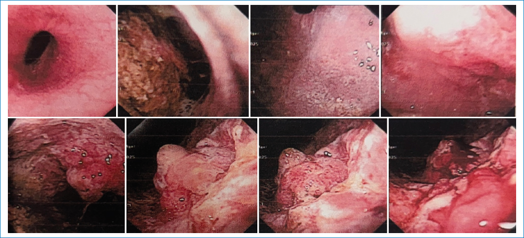
Figure 3. Upper gastrointestinal endoscopy images showing an infiltrative gastric lesion involving the antrum and pylorus, with regular mucosa, friability, and marked luminal narrowing consistent with complete pyloric stenosis. Findings are suggestive of gastric neoplasia.
Treatment was carried out by a multidisciplinary team that included hematology, oncology, surgery, gastroenterology, and nutrition. The initial treatment consisted of filgrastim for hematologic optimization, folic acid 5 mg daily, prednisone 5 mg daily doses, and nutritional watch as a result of severe malnutrition and sarcopenia. The patient underwent five cycles of the FLOT protocol treatment with poor adherence as a result of a lack of drugs and environmental determinants in the family. The main surgical management was total gastrectomy with bursectomy, splenectomy, cholecystectomy, and mechanical esophagojejunostomy with tumor resection. In the post-operative period, hematologic care, nutritional rehabilitation, and follow-up were provided. Ultimately, the patient died as a result of chronic and sustained malnutrition despite all attempts to restore his nutritional status.
Literature review
Methodology
A systematic review was conducted on PubMed and Google using the keywords: gastric adenocarcinoma and FA, including clinical cases, literature reviews, and original articles, with no restrictions on year of publication.
Forty-one articles were found, of which 32 were excluded due to the lack of association between gastric cancer and FA. Of the nine articles included in the study, three are original articles, three are case reports and literature reviews, and three are case reports only. Artificial intelligence (AI) support was used for spelling and text editing (Fig. 4).

Figure 4. Selection of articles regarding FA and gastric cancer. FA: Fanconi anemia.
Literature review
Below, we list the reviewed articles on FA and gastric adenocarcinoma published between 1981 and 2025. Table 1 lists case reports of patients with FA and gastric cancer, table 2 lists cases reports and reviewing the literature on FA and gastric cancer, and table 3 lists original articles on FA and gastric cancer (Tables 1–3).
Table 1. Case reports of FA and gastric cancer
| Case | Country | Age | FA gene/mutation | Type of gastric cancer | Key findings | Outcome/key conclusion |
|---|---|---|---|---|---|---|
| Zheng et al., 20254 | China | 32 | FANCD2 pathogenic variant (N1378Sfs*5) | Poorly differentiated gastric adenocarcinoma | Early onset, highly aggressive tumor, severe hematologic fragility | Rapid progression. Authors propose FANCD2 as a genetic risk factor |
| Pavithran et al., 20025 | India | 22 | FA diagnosed in adulthood | Intestinal-type gastric adenocarcinoma | FA diagnosed at 22, rapid progression to gastric cancer | Died 4 month later, after diagnosis. Authors recommend early endoscopic checkup |
| Hill et al., 19818 | United Kingdom | 21 | Classic FA | Gastric carcinoma | Early historical documentation of FA-associated gastric cancer | Died due to massive gastrointestinal bleeding |
|
FA: Fanconi anemia; FANCD2: Fanconi anemia complementation group D2. |
||||||
Table 2. Case reports with literature reviews of FA and gastric cancer
| Article | Country | Age | FA gene/mutation | Type of cancer | Contribution | Key conclusion |
|---|---|---|---|---|---|---|
| Huang et al., 20187 | Taiwan | 65 | FANCA D1359Y | Gastric adenocarcinoma + diffuse gastric polyposis | Reviews FA pathway alterations through gastric cancer molecular subtypes | Monoallelic FANCA variants may predispose to gastric cancer |
| Xia et al., 20206 | China | 48 | FANCA (germline heterozygous mutation) | Gastric adenocarcinoma and thyroid papillary carcinoma. | Revision of primary malignancies associated with FANCA mutations | Increased risk of multiple malignancies associated with FANCA mutation, resulting in poor prognosis |
| Dyaczyński et al., 20241 | Poland | 31 | FA | Appendiceal carcinoma (Mucinous, focal mucocellular malignancy, grade 3), lip cancer | Review highlighting the predisposition of multiple malignancies in FA patients | Emphasizes the need for intensive and early oncologic surveillance |
|
FA: Fanconi anemia; FANCA: Fanconi anemia complementation group A. |
||||||
Table 3. Original articles of FA and gastric cancer
| Article | Country | FA gene/mutation | Type of cancer | Contribution/key finding | Outcome/key conclusion |
|---|---|---|---|---|---|
| Wang et al., 20249 | China | FANCA overexpression | Gastric cancer | Experimental study, which demonstrates that FANCA promotes G1/S cell cycle progression, proliferation and invasion in gastric cancer cells | Identifies FANCA as an oncogenic driver and a potential target in the treatment of gastric cancer |
| Nepal et al., 201710 | United Kingdom | FA pathway genes (FANCA, FANCD2, BRCA axis) | Multiple cancers, including gastrointestinal tumors | Synthesis of FA signaling, DNA damage response, and carcinogenesis | Identifies FA signaling as a tumor suppressor whose disruption promotes cancer |
| Zhang and Fu, 202111 | China | ALDH2 pathway (DNA damage, associated metabolism) | Gastric and other gastrointestinal cancers | Reviews the function of ALDH2 dysfunction in aldehyde accumulation, genomic instability, and tumor progression | Positions ALDH2 as a complementary molecular pathway which contributes to gastrointestinal carcinogenesis |
|
FA: Fanconi anemia; GC: gastric cancer; FANCA: Fanconi anemia complementation group A; FANCD2: Fanconi anemia complementation group D2; BRCA axis: BRCA-related DNA repair pathway; ALDH2: aldehyde dehydrogenase 2. |
|||||
Patients with FA have consistently developed poorly differentiated gastric adenocarcinomas, frequently due to chronic gastritis, intestinal metaplasia, and polyposis, according to the clinical reports reviewed. Cases reported since the 1980s5,8 pioneered the first associations between FA and gastric cancer, while recent studies4 have pinpointed signatures of mutation, including FANCD2 N1378Sfs*5, that change the transcription of more than 30 genes associated with apoptosis and chromatin modulation.
The diagnosis of gastric cancer in patients with FA based on classic oncological approaches (endoscopy, biopsy, imaging studies) is limited by hematological frailty and poor tolerance of aggressive treatment. Early detection is critical – however, the earliest symptoms are usually non-specific (dyspepsia, early satiety, epigastric pain).
The introduction of genetic and proteomic biomarkers based on the nsSNP analysis in FANCA provides novel potential to predictive earlier risk and patient stratification. The in silico monitoring enables the detection of deleterious mutations and the association between mutations and patient susceptibility level2.
New studies12 showed that the composition of the gut microflora of gastric cancer patients underwent remarkable changes in recent years in the gut microbiome, indicating an important involvement of the microbiota in tumorigenesis and tumor progression. Bridging genomic and proteomic data in clinical practice is one of the major steps forward to precision medicine.
Functional evaluation of FANCA mutations and complementary DNA repair genes, such as FANCA and other DNA repair genes, informs the development of personalized surveillance and treatment. This work would enable pharmacologically effective therapies with reduced toxicity due to the lower toxicity of the therapeutic compounds and thus a much more complete answer to a critical and widely accepted question about gene therapy in this cancer group.
Moreover, FANCA identification of ligand binding sites represents an opportunity for target therapies that partially reinvigorate DNA repair. Such approaches may further enhance the survival and quality of life of patients with FA and gastric cancer.
AI can be used for automated systematic reviews that can synthesize large literature streams and identify potential patterns. They would then enable interdisciplinary, meta-research to integrate genetic, oncologic, and computational biology considerations13.
Nevertheless, innovation must be counterbalanced by practical implementation, given the feasibility, accessibility, and equity issues involved in the application of new diagnostic and therapeutic technologies.
Conclusions
The available data demonstrate that FA and gastric cancer share a similar biological basis in the form of DNA repair deficiency and genomic instability.
Such mutations in FANCA, FANCI, and FANCD2 not only account for hematological predisposition but also predispose the developing of aggressive solid tumors at an early stage in the life course.
Identifying deleterious mutations, generating more proteomic biomarkers, and discovering the gut microbiome provide new approaches to early identification and prevention. But obstacles persist in applying genomic data clinically and devising individualized care protocols.
The future of FA and gastric cancer research is in interdisciplinary cooperation, AI use in the routine review setting, and strategies that integrate science and patients’ health.
The patient was very young when he presented, and due to his socioeconomic conditions, it was not possible to determine the cytogenetic alterations of the tumor in time. However, it is vitally important to make recommendations for the timely detection of this type of pathology in patients with FA, given the importance of ensuring the best clinical conditions for the patient to achieve better survival outcomes.
Funding
The authors declare that they have not received funding.
Conflicts of interest
J. Loría-Castellanos is member of the editorial committee of the journal Anales Médicos. The other authors declare no conflicts of interest.
Ethical considerations
Protection of human subjects and animals. The authors declare that no experiments on humans or animals were performed for this research.
Confidentiality, informed consent, and ethical approval. This study does not involve personal patient data, medical records, or biological samples, and does not require ethical approval. SAGER guidelines do not apply.
Declaration on the use of artificial intelligence. The authors declare that no generative artificial intelligence was used in the writing or creation of the content of this manuscript.
