
Introduction
Transpedicular screws have been widely used in pathologies requiring alignment correction to maintain stability and promote adequate fusion in patients with fractures, spondylolisthesis, tumors, or scoliosis.
The most important factors affecting screw stability are: screw size, insertion depth, orientation, insertion technique, and bone mineral density1.
With the increased use of these screws, potential complications may increase, including screw malposition, infection, pseudoarthrosis, nerve irritation, and screw or rod breakage2.The presence of these complications plays an important role since, apart from poor clinical outcomes, they increase the cost related to medical care and the patient’s loss of productivity3.To minimize the risks of complications and costs derived from them, adequate pre-operative planning is essential.
The pedicle is a cylinder of bone located between the lamina and the vertebral body and has an outstanding function in the stability of these screws.
Biomechanical studies recommend that a screw should be chosen that has a diameter close to the internal pedicle diameter to achieve a good interface, a recommendation valid for vertebrae with normal bone quality4.
In studies of screw pullout resistance, the pedicle provides 60% axial resistance and 80% stiffness, which suggests that the pedicle plays an important role in vertebral biomechanics and postoperative recovery. Many academics agree that pedicle width and height are the most important anatomical parameters of this structure5.
The inner pedicle diameter is very important in screw placement to achieve safe placement and strong stabilization6. It has been observed that screw pullout forces are greater when the screw diameter fills more than 80% of the pedicle and lower when this percentage is < 80%7.
Cadaver studies have reported that plastic deformity occurs in the pedicle, and this precedes fracture when the threads are larger than the endosteal diameter or when the screw diameter is > 80% of the external cortical diameter8.
The importance of the pedicle for lumbar fixations is such that multiple studies of pedicle morphometry have been conducted in various populations to determine the appropriate diameter and angulation of screws in the lumbar region. In our country, there is only one registered study conducted on cadavers regarding pedicle morphometry.
The objective of this study is to create a database focused on the Mexican population regarding pedicle morphometry and to determine if there are differences between males and females.
Materials and methods
Retrospective study of pedicle morphometry of 60 patients, performing measurements on computed tomography (CT) scans taken between January and December 2022 were done. CT scans taken of the lumbar spine with 3 mm cuts were reviewed, using a Siemens Somatom Perspective scanner at a tertiary care hospital.
Study population
The CT scans of 60 patients were evaluated, 30 men and 30 women, aged between 20 and 70 years, who attended the outpatient clinic for lumbar pain and to whom non-surgical treatment was provided.
Patients younger than 20 years and older than 70 years, patients presenting fractures of the lumbar spine, tumors, spondylolisthesis, previous lumbar spine surgery, and vertebral deformities were excluded from this study.
Measurements
The internal and external cortical pedicle width was measured; these measurements were performed on an axial projection for both the left and right pedicles at the level of the pedicle isthmus, and the values were recorded in millimeters (Fig. 1).

Figure 1. Simple computed tomography scan showing an axial cut of L5. A: external cortical width and B: internal cortical width.
The pedicle convergence angle was measured in degrees for each pedicle from L1 to L5 (Fig. 2).
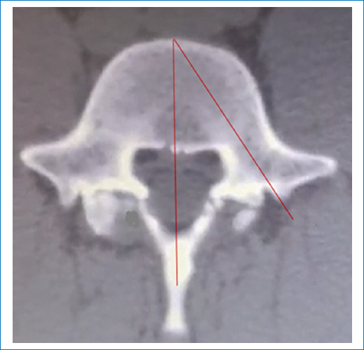
Figure 2. Simple computed tomography scan showing an axial cut of L5. The pedicle convergence angle is shown.
The internal and external cortical pedicle height was measured in a parasagittal projection for both the left and right pedicle at the level of the pedicle isthmus, and the values were recorded in millimeters (Figs. 3 and 4).
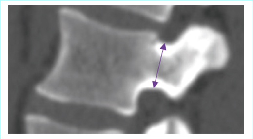
Figure 3. Simple computed tomography scan showing a parasagittal cut of L2. The arrow schematizes the external cortical height.
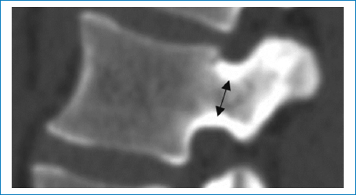
Figure 4. Simple computed tomography scan showing a parasagittal cut of L2. The arrow schematizes the internal cortical pedicle height.
Statistical analysis
The differences in these parameters in women and men were evaluated using the Mann-Whitney U statistical test, and p < 0.05 was accepted as an indication of statistical significance.
This research work complies with the ethical aspects in research matters, respects the principles of privacy, dignity, and well-being of the participants, and the anonymity of the participants was maintained at all times.
Results
The mean age was 51.7 ± 13 and 49.7 ± 12.6 years, for men and women, respectively.
At L1, the smallest parameter found in men was the internal cortical pedicle width, and the mean was 4.8 ± 2.7 and 3.67 ± 2.87 mm for right and left pedicles, respectively. The right and left pedicle convergence angles were 14.1 ± 1 and 17.4 ± 0.9°, respectively. In women, the internal cortical pedicle width was the smallest parameter found; the mean was 3.3 ± 1.8 mm for both pedicles, with a convergence angle of 11.5 ± 1 and 12.7 ± 4° for right and left pedicles, respectively. In all parameters evaluated for L1, there were no statistically significant differences (Table 1).
Table 1. Pedicle parameters of L1 vertebra
| Variables | Sex | Mean | SD | Mean difference | p |
|---|---|---|---|---|---|
| Internal cortical pedicle height_r | Men | 7.83 | 4.28 | 1.05 | 0.328 |
| Women | 6.77 | 3.27 | |||
| Internal cortical pedicle height_l | Men | 5.78 | 4.99 | 0.31 | 0.882 |
| Women | 5.47 | 3.79 | |||
| External cortical pedicle height_r | Men | 12.61 | 5.52 | 3.04 | 0.219 |
| Women | 9.56 | 5.93 | |||
| External cortical pedicle height_l | Men | 8.81 | 7.52 | −0.31 | 0.175 |
| Women | 9.12 | 5.58 | |||
| Internal cortical pedicle width_r | Men | 4.8 | 2.69 | 1.49 | 0.946 |
| Women | 3.31 | 1.86 | |||
| Internal cortical pedicle width_l | Men | 3.67 | 2.87 | 0.29 | 0.894 |
| Women | 3.38 | 1.8 | |||
| External cortical pedicle width_r | Men | 6.95 | 3.92 | 1.89 | 0.629 |
| Women | 5.05 | 2.73 | |||
| External cortical pedicle width_l | Men | 5.36 | 4.01 | −0.035 | 0.152 |
| Women | 5.4 | 2.65 | |||
| Convergence angle_r | Men | 14.1 | 10.1 | 2.6 | 0.488 |
| Women | 11.5 | 10.1 | |||
| Convergence angle_l | Men | 17.4 | 8.7 | −5.3 | 0.111 |
| Women | 12.7 | 40.1 | |||
|
A p < 0.05 was considered statistically significant. r: right; l: left; SD: standard deviation; p: probability. |
|||||
At L2, the smallest parameter found in men was the internal cortical pedicle width, and the mean was 4.9 ± 2.2 and 3.8 ± 2.7 mm in right and left pedicles, respectively. The right and left pedicle convergence angles were 15 ± 1 and 18.4 ± 0.8°, respectively. In women, the internal cortical pedicle width was the smallest parameter found; the mean was 3.1 ± 1.9 and 3.4 ± 1.8 mm in right and left pedicles, respectively, with a convergence angle of 16.9 ± 0.9 and 18.4 ± 0.8° for right and left pedicles, respectively. In all parameters evaluated for L2, there were no statistically significant differences (Table 2).
Table 2. Pedicle parameters of L2 vertebra
| Variables | Sex | Mean | SD | Mean difference | p |
|---|---|---|---|---|---|
| Internal cortical pedicle height_r | Men | 7.29 | 3.52 | 2.36 | 0.372 |
| Women | 4.93 | 3.42 | |||
| Internal cortical pedicle height_l | Men | 5.04 | 4.15 | 0.69 | 0.174 |
| Women | 4.34 | 3.73 | |||
| External cortical pedicle height_r | Men | 11.58 | 5.99 | 2.07 | 0.25 |
| Women | 9.5 | 4.92 | |||
| External cortical pedicle height_l | Men | 11.61 | 6.18 | 3.45 | 0.96 |
| Women | 8.15 | 5.69 | |||
| Internal cortical pedicle width_r | Men | 4.92 | 2.19 | 1.82 | 0.249 |
| Women | 3.09 | 1.98 | |||
| Internal cortical pedicle width_l | Men | 3.78 | 2.69 | 0.4 | 0.735 |
| Women | 3.38 | 1.77 | |||
| External cortical pedicle width_r | Men | 7.77 | 2.66 | 2.66 | 0.166 |
| Women | 5.11 | 2.74 | |||
| External cortical pedicle width_l | Men | 6.78 | 3.29 | 1.71 | 0.213 |
| Women | 5.07 | 2.76 | |||
| Convergence angle_r | Men | 15 | 1 | −1.9 | 0.274 |
| Women | 16.9 | 9.1 | |||
| Convergence angle_l | Men | 18.8 | 7.7 | 0.43 | 0.788 |
| Women | 18.4 | 8.2 | |||
|
A p < 0.05 was considered statistically significant. r: right; l: left; SD: standard deviation; p: probability. |
|||||
At L3, the smallest parameter found in men was the internal cortical pedicle width, and the mean was 5.47 ± 2.98 and 5.5 ± 3.06 mm in right and left pedicles, respectively. The right and left pedicle convergence angles were 16.5 ± 1.09 and 19.5 ± 0.97°, respectively. In women, the internal cortical pedicle width was the smallest parameter found, the mean was 4.1 ± 2 and 4.03 ± 2.38 mm in right and left pedicles, respectively, with a convergence angle of 16.5 ± 1 and 19.5 ± 0.9° for right and left pedicles, respectively. In all parameters evaluated for L3, there were no statistically significant differences (Table 3).
Table 3. Pedicle parameters of L3 vertebra
| Variables | Sex | Mean | SD | Mean difference | p |
|---|---|---|---|---|---|
| Internal cortical pedicle height_r | Men | 7.7 | 3.47 | 1.68 | 0.241 |
| Women | 6.02 | 3.29 | |||
| Internal cortical pedicle height_l | Men | 4.94 | 4.19 | 0.23 | 0.785 |
| Women | 4.71 | 3.57 | |||
| External cortical pedicle height_r | Men | 9.87 | 6.41 | −0.57 | 0.257 |
| Women | 10.45 | 4.33 | |||
| External cortical pedicle height_l | Men | 7.61 | 7.07 | −3.16 | 0.517 |
| Women | 10.78 | 4.11 | |||
| Internal cortical pedicle width_r | Men | 5.47 | 2.98 | 1.36 | 0.528 |
| Women | 4.1 | 2 | |||
| Internal cortical pedicle width_l | Men | 5.5 | 3.06 | 1.47 | 0.668 |
| Women | 4.03 | 2.38 | |||
| External cortical pedicle width_r | Men | 7.89 | 4.14 | 0.98 | 0.08 |
| Women | 6.91 | 1.88 | |||
| External cortical pedicle width_l | Men | 8.13 | 4.36 | 1.21 | 0.834 |
| Women | 6.92 | 2.44 | |||
| Convergence angle_r | Men | 16.5 | 1.09 | 0 | 0.436 |
| Women | 16.5 | 1.09 | |||
| Convergence angle_l | Men | 19.5 | 0.97 | 0 | 0.199 |
| Women | 19.5 | 0.95 | |||
|
A p < 0.05 was considered statistically significant. r: right; l: left; SD: standard deviation; p: probability. |
|||||
At L4, the smallest parameter found in men was the internal cortical pedicle height, and the mean was 7.11 ± 3.77 and 3.66 ± 4.07 mm in right and left pedicles, respectively. The right and left pedicle convergence angles were 17.5 ± 1.25 and 20.8 ± 1.12°, respectively. In women, the internal cortical pedicle width was the smallest parameter found; the mean was 4.59 ± 2.52 and 4.55 ± 2.84 mm in right and left pedicles, respectively, with a convergence angle of 21.4 ± 1.15 and 16.2 ± 1.28° for right and left pedicles, respectively. There were statistically significant differences for the internal cortical pedicle height; in the rest of the evaluated parameters, there were no significant differences (Table 4).
Table 4. Pedicle parameters of L4 vertebra
| Variables | Sex | Mean | SD | Mean difference | p |
|---|---|---|---|---|---|
| Internal cortical pedicle height_r | Men | 7.11 | 3.77 | 0.91 | 0.635 |
| Women | 6.19 | 2.48 | |||
| Internal cortical pedicle height_l | Men | 3.66 | 4.07 | −1.13 | 0.018 |
| Women | 4.79 | 3.28 | |||
| External cortical pedicle height_r | Men | 9.94 | 5.48 | 0.9 | 0.331 |
| Women | 9.04 | 4.21 | |||
| External cortical pedicle height_l | Men | 8.45 | 6.18 | −0.21 | 0.953 |
| Women | 8.67 | 4.59 | |||
| Internal cortical pedicle width_r | Men | 6.39 | 2.99 | 1.8 | 0.141 |
| Women | 4.59 | 2.52 | |||
| Internal cortical pedicle width_l | Men | 4.98 | 3.92 | 0.42 | 0.435 |
| Women | 4.55 | 2.84 | |||
| External cortical pedicle width_r | Men | 9.22 | 4.06 | 2.93 | 0.084 |
| Women | 6.28 | 4.01 | |||
| External cortical pedicle width_l | Men | 9.69 | 3.67 | 2.2 | 0.679 |
| Women | 7.48 | 3.45 | |||
| Convergence angle_r | Men | 17.5 | 1.25 | −3.8 | 0.825 |
| Women | 21.4 | 1.15 | |||
| Convergence angle_l | Men | 20.8 | 1.12 | 4.5 | 0.764 |
| Women | 16.2 | 1.28 | |||
|
A p < 0.05 was considered statistically significant. r: right; l: left; SD: standard deviation; p: probability. |
|||||
At L5, the smallest parameter found in men was the internal cortical pedicle height, and the mean was 6.52 ± 4.21 and 6.65 ± 5.03 mm in right and left pedicles, respectively. The right and left pedicle convergence angles were 17.7 ± 1.49 and 19 ± 1.53°, respectively. In women, the internal cortical pedicle height was the smallest parameter found, the mean was 5.22 ± 3.04 and 4.76 ± 3.27 mm in right and left pedicles, respectively, with a convergence angle of 30.6 ± 5.7 and 28.5 ± 1.31° for right and left pedicles, respectively. There were statistically significant differences for the internal cortical pedicle width; in the rest of the evaluated parameters, there were no significant differences (Table 5).
Table 5. Pedicle parameters of L5 vertebra
| Variables | Sex | Mean | SD | Mean difference | p |
|---|---|---|---|---|---|
| Internal cortical pedicle height_r | Men | 6.52 | 4.21 | 1.29 | 0.361 |
| Women | 5.22 | 3.04 | |||
| Internal cortical pedicle height_l | Men | 6.65 | 5.03 | 1.89 | 0.386 |
| Women | 4.76 | 3.27 | |||
| External cortical pedicle height_r | Men | 9.65 | 6.56 | 1.16 | 0.153 |
| Women | 8.49 | 4.31 | |||
| External cortical pedicle height_l | Men | 8.68 | 7.05 | 0.76 | 0.451 |
| Women | 7.92 | 4.54 | |||
| Internal cortical pedicle width_r | Men | 6.77 | 4.71 | 0.36 | 0.453 |
| Women | 6.4 | 3.45 | |||
| Internal cortical pedicle width_l | Men | 8.78 | 2.92 | 2.4 | 0.011 |
| Women | 6.38 | 3.75 | |||
| External cortical pedicle width_r | Men | 10.06 | 5.53 | 0.71 | 0.861 |
| Women | 9.34 | 5.09 | |||
| External cortical pedicle width_l | Men | 10.02 | 6.01 | −0.07 | 0.524 |
| Women | 10.1 | 4.38 | |||
| Convergence angle_r | Men | 17.7 | 1.49 | −12.8 | 0.431 |
| Women | 30.6 | 5.7 | |||
| Convergence angle_l | Men | 19 | 1.53 | −9.5 | 0.706 |
| Women | 28.5 | 1.31 | |||
|
A p < 0.05 was considered statistically significant. r: right; l: left; SD: standard deviation; p: probability. |
|||||
The mean of the internal and external pedicle width and the convergence angle gradually increased from L1 to L5 in both men and women.
Discussion
In the present study, emphasis was placed on the internal and external cortical diameters of the pedicle isthmus and on the pedicle convergence angle.
Based on the results obtained regarding the internal cortical pedicle width for L1 and L2 in males, it is possible to safely place screws with a diameter of 5.0 mm, and in the pedicles of L3, the use of 5.5 mm screws would be allowed; in pedicles of L4 and L5, the placement of screws with a diameter of 5.5 mm would be safe and up to 6.5 mm with a higher risk of plastic deformity of the pedicle.
In women, regarding the internal pedicle width, it is observed that the placement of screws with a diameter of 4.0 mm is safe from L1 to L3, with a risk of presenting plastic deformity in L1 and L2 despite the small diameter of the screw; for vertebrae L4 and L5, the safe screw diameter would be 5.0 mm.
With the safe measurements found for the Mexican population, the recommendation of Acharya et al.9 is agreed upon, who in their study concluded that the use of 7 mm diameter screws in upper lumbar levels is not justified because it can generate violation of the pedicle isthmus and lead to neurological complications.
Regarding the internal cortical pedicle width, values lower than those reported by Olmos-Alfonso et al.10 in the Spanish population were found. While the values reported by Li et al. were similar compared to this study, as were those reported by Zhe-Heng et al.11,12 The results obtained for external pedicle width by Urrutia-Vega et al.13 in their study conducted on 60 cadavers are similar to the values found in men; the results of this study in women were lower.
The measurements of the internal and external pedicle width showed a gradual increase from L1, with the values obtained at L5 being higher for women and men; this trend was similar to that found by Sugisaki et al14.
In the results obtained for the internal and external pedicle height, a gradual decrease was found, being higher at L1 and lower at L5, in both men and women. These results contrast with those found by Torun et al.15, who reported that these values practically remain constant in all lumbar vertebrae in men and women. The results in these parameters were similar to those reported by Cook and Baker16 in a Maori population, where pedicle height values decreased gradually, being higher at L1 and lower at L5.
The measurements obtained for the external cortical pedicle height in males were similar to those reported by Singel et al. in Gujarat, India. The results in women for this same parameter were similar to those reported by Verma and Agrawal in their study on dry lumbar specimens17.
Regarding the pedicle convergence angle, a gradual increase was found; the angle was smaller at L1 and larger at L5; this was true for both women and men. This trend was similar to that reported by Chen et al.18 and by Zindrick et al.19, in Chinese volunteers and in their cadaver study, respectively.
In this population sample, the pedicle convergence angle was greater at L5, but these values were lower than those reported by Grivas et al.20 in a Greek population.
Conclusion
This study provides a database on pedicle morphometry specific to the Mexican population sample. The mean values obtained will help us to adequately select the diameter of the transpedicular screw required to be applied for each lumbar vertebra. In the population studied, 5.0 mm diameter screws are safe for pedicles of vertebrae L1 and L2, and 5.5 mm screws are safe for placement from L3 to L5 in men. In women, it is safe to use screws with a diameter of 5.0 mm at L4 and L5; while from L1 to L3, the safe diameter is 4.0 mm, with an increase in plastic deformity in these vertebrae.
Funding
The authors declare that they have not received funding.
Conflicts of interest
J. Loría-Castellanos is a member of the editorial committee of the journal Anales Médicos. The other authors declare no conflicts of interest.
Ethical considerations
Protection of human subjects and animals. The authors declare that no experiments on humans or animals were performed for this research.
Confidentiality, informed consent, and ethical approval. The authors have obtained approval from the Ethics Committee for the analysis of routinely collected and anonymized clinical data; therefore, individual informed consent was not required. Relevant ethical recommendations have been followed.
Declaration on the use of artificial intelligence. The authors declare that no generative artificial intelligence was used in the writing or creation of the content of this manuscript.
